What is lumbar laminectomy?

Lumbar laminectomy or decompression is a microsurgical procedure that allows a part of the posterior spinal column (lamina) to be resected unilaterally (hemilaminectomy) or bilaterally (laminectomy) to free the spinal canal and its internal neural elements (spinal cord, nerves, and nerve roots).

What cases is it used for?

It is used in cases of spinal degenerative pathologies. The most frequent pathology that needs treatment through this procedure is spinal stenosis, a narrowing of the spine that affects the spinal cord and nerves.

This procedure can also be used for foraminal recess stenosis, either secondary or not to a herniated disc, osteoarthritis of the disc and/or posterior joints, spondylopathies, and occasionally degenerative spondylolisthesis

It can also be performed as an added procedure during a lumbar fusion to complete the treatment.

What does the procedure involve?

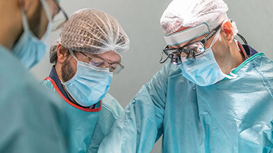
The surgery is done under general anesthesia with the patient in a face-down position on the operating table. 

First, a medium length incision is made in the skin, at the level to be treated (for example, L4-L5 or L5-S1). After separating the musculature, we reach the bone structures. The layer to be treated is exposed and held with a surgical separator. At this point the surgical microscope is introduced into the operative field.

With the help of a tool called a high-speed motor, the surgeon mills a part of the lamina (hemilaminectomy, one side only) or the entire lamina (laminectomy). In the second case, the resection of another element of the posterior column called the spinous process is performed, which is a more invasive surgical procedure as it involves separation of the musculature from both sides.

There is a less invasive variant of the laminectomy technique that consists of a bilateral resection through a unilateral access (over the top), which allows the laminas to be resected from both sides, separating the musculature on one side only, and which is minimally invasive, faster and equally effective in the long term.

After removing the lamina, a ligament underneath it, called a yellow ligament because of its straw-like color, is resected. This ligament is strongly anchored to the lamina and in degenerative pathologies contributes to compression of the neural elements (medulla, nerves and nerve roots).

By resection of the laminas and yellow ligament, we achieve the release (decompression) of the neural elements. Sometimes, it is also necessary to physically mobilize the nerves and roots that may be blocked and attached to bone structures due to the pathology. If there is a herniated disc, it is removed during the same operation.


The advantages of this technique, when it is appropriate for the specific patient’s case, is that it avoids the destabilization of the spine, achieves the decompression of the neural elements, conserves the patient's physiological mobility, and there is no need to use prosthetic material (screws, interbody cages, implants, etc.).

In cases where the hemilaminectomy/laminectomy is performed in association with another surgical procedure, such as a fusion then an arthrectomy (a wide resection including the posterior joints), will be performed.

Recovery and rehabilitation after a lumbar laminectomy

When there are no complications, the recovery from this surgery is quite fast. Usually, the patient is standing and walking the day after surgery. If the pain is controlled sufficiently with oral medication, the patient may be discharged to return home 1 or 2 days after the surgery.

During the first 4/6 weeks post-surgery, the patient should take it easy. This means he can and should take short walks of 5-15 minutes several times a day, but must avoid bending over forwards, carrying weight, physical exercise, and should not yet return to work. 

About 7 to 10 days after the operation, the patient will see a member of the nursing staff at Instituto Clavel to check how the surgical wound is healing and to have the stitches or staples removed from the suture.

Between 4 and 6 weeks after surgery, the patient will have a follow-up visit with a neurosurgeon who will assess his general condition and clinical evolution. If everything is going well, the patient can gradually return to his usual activities 6 weeks after the operation. At 10 to 12 weeks after surgery, if there are no contraindications, the patient can gradually start doing physical activity and exercise. 

At the beginning of the return to physical activity, we recommend avoiding impact or contact sports, or activity that involves flexing and twisting. Then, later on, the patient can gradually return to whatever physical and exercise activity they like, because with this kind of surgery, there is no loss of physiological mobility.

All this means that, it would be good to start with activities like elliptical or stationary bike, swimming, Nordic walking, etc., and leave sports such as tennis, paddle tennis, soccer and golf for later on.

Remember, that as a general recommendation, even if you have not had surgery, it is very important to warm up before and stretch after doing any exercise.

Although the use of a lumbar support is not mandatory after this surgery, it can be worn during the first two weeks after the operation to facilitate tissue healing. Later, it can be used in case of physical activity or going in any means of transportation.

Risks of lumbar laminectomy

There are very few risks associated with a lumbar laminectomy. However, as with any surgery, the possibility of general complications related to anesthesia must be taken into account, the possibility of either deep or superficial infection of the surgical wound, and the possibility of post-surgical hematoma (bleeding after surgery), which may require a new surgery. 

Regarding the specific risks, it is necessary to mention the possibility of an injury to the dura mater (the layer that covers the medulla and nerves) with result of leakage of a liquid called cerebrospinal fluid (CSF). If this occurs, the lesion is repaired and sealed, with biological patches and/or biological glues if necessary. It is very important that this liquid not leak through the skin as this could lead to wound infection. In the event of this complication, the patient will be asked to remain in bed for 24-48 hours in order for the scar to heal. It rarely requires new surgery

Another, much rarer, possible complication of this surgery is a worsening of the neurological injury that was present before surgery, or the appearance of a new neurological lesion. When this occurs, it is due to manipulation of the nerves during surgery and is usually a transient complication, rarely permanent, and it usually improves with medication and rehabilitation

Who are the doctors at Instituto Clavel who perform lumbar laminectomy?

Sources

  • Outcome of unilateral versus standard open midline approach for bilateral decompression in lumbar spinal stenosis: is "over the top" really better? A Swiss prospective multicenter cohort study. Ulrich NH, Burgstaller JM, Gravestock I, Pichierri G, Wertli MM, Steurer J, Farshad M, Porchet F.J Neurosurg Spine. 2019 Apr 26:1-10. doi: 10.3171/2019.2.SPINE181309. [Epub ahead of print] 
  • Minimally Invasive Approaches for Surgical Treatment of Lumbar Spondylolisthesis. Hussain I, Kirnaz S, Wibawa G, Wipplinger C, Härtl R. Neurosurg Clin N Am. 2019 Jul;30(3):305-312. doi: 10.1016/j.nec.2019.02.004. Epub 2019 Apr 19. Review.
  • Microdecompression for lumbar spinal canal stenosis. Weiner BK, Walker M, Brower RS, McCulloch JA. Spine (Phila Pa 1976). 1999 Nov 1;24(21):2268-72.
  • Relief of lumbar canal stenosis using multilevel subarticular fenestrations as an alternative to wide laminectomy: preliminary report. Young S, Veerapen R, O'Laoire SA.Neurosurgery. 1988 Nov;23(5):628-33.
  • Outcome after less-invasive decompression of lumbar spinal stenosis: a randomized comparison of unilateral laminotomy, bilateral laminotomy, and laminectomy. Thomé C, Zevgaridis D, Leheta O, Bäzner H, Pöckler-Schöniger C, Wöhrle J, Schmiedek P.J Neurosurg Spine. 2005 Aug;3(2):129-41.
  • Rehabilitation after lumbar disc surgery. Oosterhuis T et al. Cochrane Database Syst Rev. (2014)
     

Want to know more?
Tell us about your case!

Contact us so that we can give you a personalized assessment.

Share on: