Cervical laminectomy or decompression is a surgical procedure that allows a part of the posterior spine to be removed to decompress the spinal cord and nerves.
Acabar con el dolor es posible y nuestro equipo quiere ayudarle a conseguirlo. Dé el primer paso contactando con nosotros.
Madrid: +34 919 148 441
Barcelona: +34 936 090 777
Llevamos la excelencia neuroquirúrgica a Sevilla, ofreciendo servicios especializados en neurocirugía de columna y cráneo.
¡Llama al +34 955 277 751 o +34 633 143 686 para más información!
Cervical laminectomy or decompression is a surgical procedure that allows a part of the posterior spine to be removed to decompress the spinal cord and nerves.

It is used in cases of degenerative pathologies of the cervical spine that cause a narrowing of the cervical spinal canal and spinal compression (cervical spondyloarthrosis with myelopathy or spinal distress).
This procedure is indicated particularly when cervical disc degeneration is accompanied by calcification of the posterior longitudinal ligament. It is also performed in cases of medullar tumor pathology, vascular malformation pathology or in surgical correction of prior failed surgery.
The surgery is done under general anesthesia with the patient in a face-down position on the operating table. Intraoperative neurophysiological monitoring is used throughout, to monitor the medulla and nerves during the surgery.
The patient's head is usually immobilized by attaching a specific device called a Mayfield so that the neck does not move during surgery. A medium length incision is made in the skin, usually from the lower part of the head to the upper dorsal part (10-12cm). After separating the musculature, we reach the bone structures. The layers to be treated are exposed, bilaterally and held with a surgical separator.
The posterior bone structures (spines and laminas) are removed with the help of tools called gouges and kerrisons. After removing the laminas, a ligament underneath it, called a yellow ligament because of its straw-like color, is resected. This ligament is strongly anchored to the lamina and in degenerative pathologies contributes to. compression of the neural elements (medulla, nerves and nerve roots).
By resection of the laminas and yellow ligament, we achieve the release (decompression) of the neural elements. In this surgery on the cervical spine, the posterior decompression technique using laminectomy creates instability of the cervical spine. Over the years, experience has taught us that, not only did this lead to a problem of instability, but also led to deformity of the spine (cervical kyphosis). For these reasons, in current practice, the cervical spine is fixed with posterior screws and rods.

In the absence of any complications, recovery from this surgery is quite fast. The patient usually has a drain that is removed within 24-48 hours. The patient will begin walking the day after surgery. Depending on how the level of post-surgical pain evolves, the patient can be discharged 4-6 days after surgery.
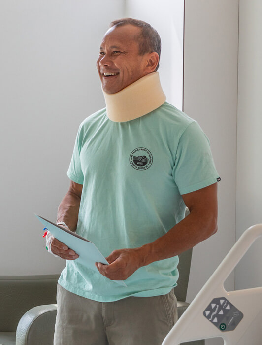
During the first 4 or 6 weeks after surgery, the patient should take it easy, this means that he can and should walk, for about 5-15 minutes frequently throughout the day. The patient should wear a rigid collar for the first 4 weeks, although it can be removed when sitting or in bed, after that, it can be replaced with a soft collar for an additional 2 weeks. At this time the patient should avoid bending his neck, carrying weight, or doing physical exercise, and should not yet go back to work.
Next, about 7 to 10 days after the operation, the patient will see a member of the nursing staff at Instituto Clavel to check how the surgical wound is healing and to have the stitches or staples removed from the suture
Between 4 and 6 weeks after surgery, the patient will have a follow-up visit with a spine specialist who will assess his general condition and clinical evolution. If everything is going well, the patient can gradually return to his usual activities 6 weeks after the operation.
Once the stability of the implants has been confirmed (usually between 3-6 months following surgery), and if there are no contraindications, the patient can gradually start doing physical activity and exercise, although contact sports and impact sports should be limited and only after it has been confirmed that the fusion is complete (about 1 year after surgery). During the entire recovery process, the patient will receive all the support they need, both in person and remotely, as part of our Preparation, Empowerment, and Recovery (PER) program.
As with all surgery, the possibility of general complications related to anesthesia must be taken into account, the possibility of either deep or superficial infection of the surgical wound, and the possibility of post-surgical hematoma (bleeding after surgery), which may require a new surgery
Due to the sudden decompression of the medulla, in the case of myelopathy, sometimes there may be worsening of the symptoms the patient was having prior to surgery, and in this case, further rehabilitation will be necessary.
Regarding the placement of the implant material (screws), it is important to note that in some cases, the material may not fuse with the bone (pseudo-arthrosis) and corrective surgery will be needed. Vertebral artery injuries are very rare (less than 2%), as well as nerve (1%) or root injuries (C5 paresis, difficulty lifting the shoulder).
Contact us so that we can give you a personalized assessment.